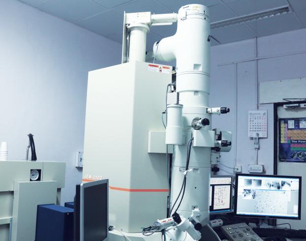
Cryogenic Transmission Electron Microscopy
Cryo-TEM involves performing Transmission Electron Microscopy TEM analysis while keeping the sample at cryogenic temperatures, i.e. -170°C (or 103 K).
Transmission Electron Microscopy (TEM) is a technique that images a sample using an electron beam. High energy electrons (80-200 keV) are transmitted through electron transparent samples (~100 nm thick) and imaged on a plane.
The main reasons for the use of low temperature are
The study of thin, frozen slices of suspensions, allowing for morphology studies of particles in their dispersed state.
The reduction of sample heating by the electron beam and thus the reduction of potential beam damage of sensitive materials

The study of low-temperature phases of crystalline materials
The production of thin (~100 nm) slices of frozen aqueous dispersions (‘vitrified ice layers’) is achieved using a Vitrobot ™ sample preparation tool, a dedicated cryo-sample-transfer unit and a cryo-TEM sample holder.
Ideal Uses of Cryo-TEM
- Colloidal dispersions of liposomes, polymersomes, emulsions
- Studies of particle clustering in the dispersion
- Electron-beam sensitive samples
- Low-temperature crystal phase studies
Strengths
- Allows for imaging of soft matter in its near natural state in an aqueous dispersion
- Allows for imaging of otherwise too beam sensitive materials
Limitations
- The sample preparation of aqueous dispersions requires method development for different sample types, as it is very dependent on the particle concentration, particle size and material viscosity
- The sample thickness of vitrified ice layers is determined by the particle size. The transparency of the sample thus sets a limit to the particle size at a few hundred nm
- The analysis time for Energy Dispersive X-ray Spectroscopy (EDS) analysis is limited because of beam-induced sublimation of the frozen water layer, even at cryogenic temperatures
Cryo-TEM Technical Specifications
- Signals Detected: Transmitted electrons
- Elements Detected: B-U (with EDS)
- Detection Limits: 1 at%
- Imaging/Mapping: Yes (with EDS)
